Research News
Discovery of Two Types of Vesicle Fusion Mechanisms via Visualization of Vesicle Movement in Living Cells
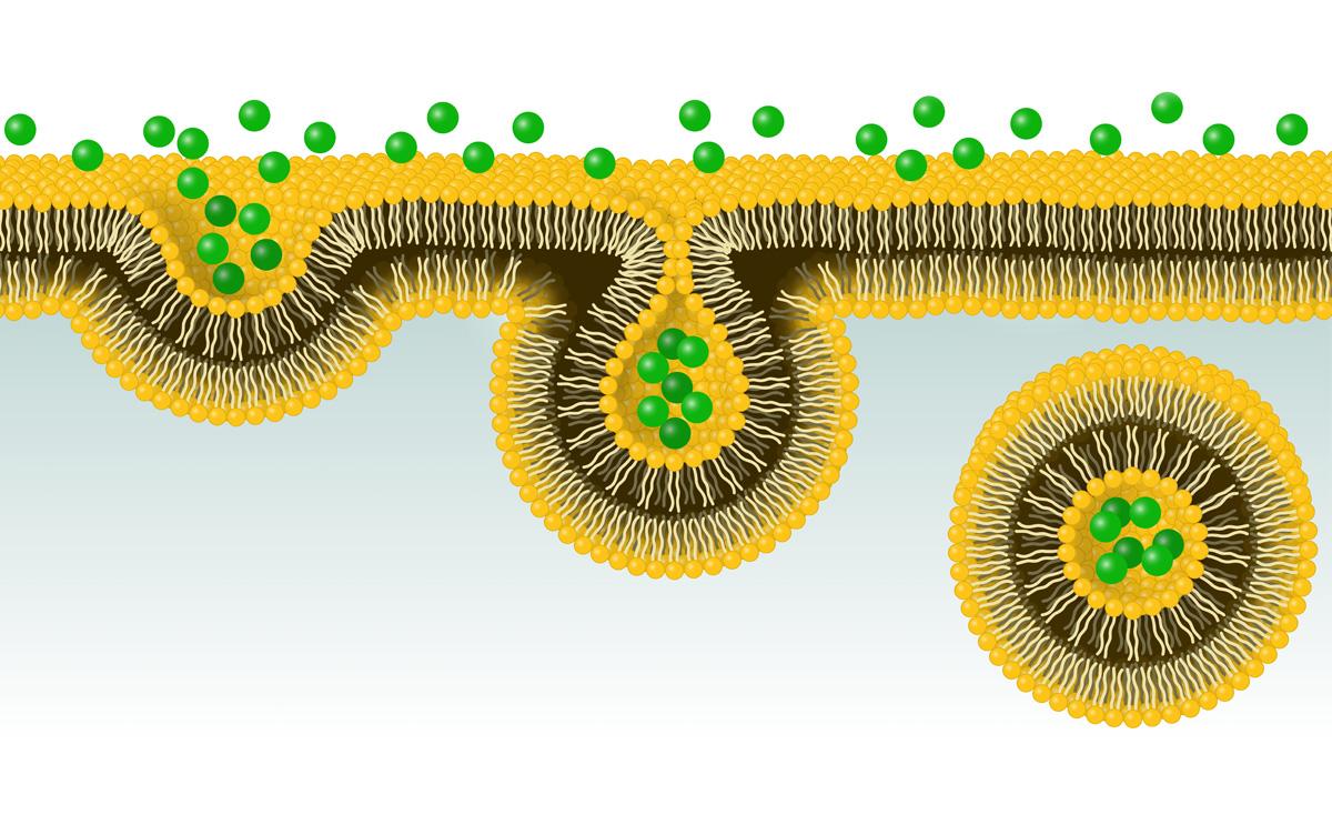 Image by J. Marini/Shutterstock
Researchers at University of Tsukuba developed a novel technique to visualize the vesicle fusion process using yolk sac cells encasing mouse embryos. By labeling vesicles, which transport substances within cells, with fluorescent substances, researchers discovered two different modes of vesicle fusion and the role of cytoskeletal actin in regulating the fusion process.
Tsukuba, Japan—Cells intake substances from the outside world by encapsulating them in vesicles called endosomes, which are subsequently transported throughout the cell. During the transport process, vesicles fuse with other intracellular organelles. However, observing this process is challenging in many cells due to their small size and the complexity of underlying regulatory mechanisms.
In this study, the researchers focused on the outermost visceral endoderm cells of mouse embryos. By labeling endosomes with fluorescent markers, they successfully observed the fusion process under a microscope. They identified two distinct modes of endosomal fusion: homotypic fusion, where two endosomes fuse rapidly to form a single vesicle, and heterotypic fusion, in which lysosomes slowly absorb endosomes. Further, a mathematical analysis of the vesicle fusion process using a mechanical model of the vesicle membrane revealed that the vesicle size determines the fusion mode: homotypic fusion occurs in small vesicles and heterotypic fusion happens in large ones. Moreover, the application of fluctuating forces to the vesicle membrane facilitated homotypic fusion even in large vesicles. The binding of endosomes to the cytoskeletal protein called actin, which is believed to generate these fluctuating forces, appears to promote homotypic fusion. Furthermore, cofilin, which promotes actin turnover, and myosin, which interacts with actin, were crucial for vesicle fusion. Using this observation system, the researchers expect to elucidate the regulatory mechanisms controlling the processes of intracellular vesicle fusion and trafficking.
###
This work was supported in part by KAKENHI (17024006) and the 21st Century COE program from the MEXT, Japan, KAKENHI (JP22H04926), Grant-in-Aid for Transformative Research Areas - Platforms for Advanced Technologies and Research Resources "Advanced Bioimaging Support" from JSPS, Japan, and Takeda Science Foundation.
Original Paper
- Title of original paper:
- Actin dynamics switches two distinct modes of endosomal fusion in yolk sac visceral endoderm cells
- Journal:
- eLife
- DOI:
- 10.7554/eLife.95999
Correspondence
Professor MASU Masayuki
Institute of Medicine, University of Tsukuba
Assistant Professor KOIKE Seiichi
Laboratory of Molecular and Cellular Biology, Graduate School of Science and Engineering for Research, University of Toyama
Associate Professor TACHIKAWA Masashi
Graduate School of Nanobioscience, Yokohama City University
Professor NEMOTO Tomomi
Exploratory Research Center on Life and Living Systems (ExCELLS) / National Institute for Physiological Sciences, National Institutes of Natural Sciences
Related Link
